


Osteoarthritis of the hip joint or coxarthrosis is a chronic and slow degenerative process in the joint of the femoral head and acetabulum of the pelvic bone. With this disease, bone and cartilaginous tissues are deformed, and as it progresses, it leads to significant limitation of leg movements and disability. All components of the joint are involved in the process: bones, joint capsules that cover them, cartilage, ligaments, muscles. Symptoms and treatment of osteoarthritis of the hip joint (coxarthrosis) vary from person to person; the disease usually occurs in middle-aged and elderly people, although such changes can develop after 20 years.
The main signs of arthrosis of the hip joint (coxarthrosis) are pain and stiffness of movements. Very often, its development is preceded by lesions and joint pathologies of an inflammatory and non-inflammatory nature. Coxarthrosis is one of the most common arthrosis, associated with a significant load on the hip joint.
In its development, the disease goes through several stages. In the initial stages, coxarthrosis can be treated conservatively, but as the process progresses only surgical treatment is effective. Therefore, you should not delay visiting a specialist and sign up for a consultation. In clinics you can undergo examinations and receive conservative treatment.
Causes
Coxarthrosis of the hip joint can be primary or secondary, that is, arise against the background of any disease or injury of the musculoskeletal system. Let us consider in more detail the factors influencing the development or leading to coxarthrosis of the hip joint.
- Exogenous- these are environmental factors: heavy physical activity, consequences of serious injuries - fractures, dislocations, ligament tears, unfavorable working conditions associated with heavy lifting, prolonged sitting.
- Endogenous— these are chronic infectious-inflammatory and autoimmune diseases: rheumatoid, reactive, psoriatic arthritis. As well as metabolic disorders: gout, diabetes.
- Congenital diseases.Dysplasia (impairment of joint formation) and osteochondropathy (malnutrition of joint structures resulting in necrosis, bone destruction) can also lead to coxarthrosis. For example, congenital dislocation of the hip, aseptic necrosis of the femoral head - Perthes disease.
- Genetic predispositionoften causes coxarthrosis of the hip joints. This includes a mutation in the procollagen type II gene.
- Elderly age.Most often, the development of coxarthrosis of the hip joint is due to inevitable age-related changes.
- Flooring. It is believed that arthrosis occurs more often in women than in men. This is due to the influence of the female sex hormones, estrogen, on mineral metabolism and bone density.
- Excess body weight.There is a direct relationship between excess body weight and the onset of osteoarthritis. The greater the body weight, the greater the likelihood of developing arthrosis of the hip joint, since excess adipose tissue increases the load on the joints, and adipose tissue produces pro-inflammatory substances that damage cartilage tissue.
- Professional sportscan cause the development of coxarthrosis due to excessive stress on the joints and frequent injuries. Potentially dangerous sports include weightlifting, skydiving and acrobatics.
Under the influence of these factors, changes gradually occur in the joint cavity at the cellular level: decay processes begin to prevail over synthesis processes, metabolism changes, the volume of joint fluid nourishing the cartilage tissue decreases, and the cartilage it becomes thinner. As a result, the joint "dries out" and decreases in volume. Along the edges of the articular surfaces of the bones, bone growths appear - osteophytes, which reduce the range of motion of the joint and thus reduce the load on it.
Symptoms
How quickly does osteoarthritis of the hip joint (coxarthrosis) develop? Symptoms increase gradually, and in the early stages a person may not pay due attention to them and consider them fatigue. This is dangerous because it is at the beginning of the degenerative process that the treatment has the greatest effect.
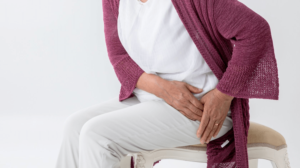
The first clinical symptoms of coxarthrosis are pain and limited mobility caused by muscle spasm.
The pain can vary in intensity and duration. Initially, unpleasant sensations are mild and short-lived. The provoking factor for their appearance is prolonged walking or intense physical activity.
Limitation of joint mobility occurs due to severe pain. The patient's gait changes: the buttocks protrude backwards, the body leans forward when transferring weight to the injured side, and the person limps.
Swelling in the joint area, which is usually invisible due to the muscle and fat layer, creaking of the joints during movement, functional shortening of the lower limb is also possible.
The presence of some signs and their severity depend on the stage of coxarthrosis. There are 4 clinical and diagnostic stages of coxarthrosis, determined by the degree of damage to the articular cartilage:
- Coxarthrosis 1st degreecharacterized by asymptomatic or periodic pain that occurs only after intense physical activity, such as running or long walking. The pain is localized in the joint area, less often it spreads to the entire thigh and even the knee. After rest it usually disappears. On the x-ray of the hip joint, no changes are noted or a slight narrowing of the joint space is observed. Magnetic resonance imaging reveals signs of cartilaginous tissue heterogeneity.
- For 2 degree coxarthrosisthe pain becomes more intense, appears with little physical activity, and sometimes at rest, and can radiate to the thigh and groin area. Lameness appears after significant physical exertion. The range of motion of the joint decreases: abduction and inward rotation of the hip are limited. X-ray photographs reveal obvious irregular narrowing of the joint space and isolated osteophytes (growths of bone tissue) along the edge of the glenoid cavity. An MRI at stage 2 of coxarthrosis reveals obvious erosions and cracks of the cartilage with its thinning less than half.
- For 3rd degree coxarthrosisthe pain becomes constant and often disturbs patients during sleep. Walking is difficult, which forces the patient to take a forced position of the body, relying on a healthy leg or a stick. The range of motion of the joint is severely limited. In the radiographs the joint space is practically absent and numerous osteophytes have formed on the bone surfaces. MRI shows destruction of more than half of the volume of cartilaginous tissue. However, the third stage can still be treated conservatively.
- Stage 4 osteoarthritis of the hip joint (coxarthrosis)characterized by a significant loss of joint function. The whole leg hurts: joint, groin, gluteal region, hip, knee, ankle. Flat feet develop, the leg shortens and its muscles atrophy. On x-ray: numerous large osteophytes, joint space is absent or reduced to a minimum. Stage 4 is not amenable to conservative treatment; hip replacement is performed. The operation reduces pain, improves the functioning of the leg and the patient's quality of life.
Diagnosis of arthrosis of the hip joint
The basis for diagnosing arthrosis of the hip joint is an initial consultation with a specialist. The doctor clarifies the disorders: where the pain is located, when and why it occurs, where it goes, what reduces and intensifies it, what causes it. Then a visual inspection, palpation, gait assessment is required, and special tests are performed to detect joint dysfunction.
The diagnosis of coxarthrosis is made on the basis of clinical signs and data from further instrumental studies, the main of which is radiography of the joint. There are no characteristic laboratory signs for the diagnosis of osteoarthritis, however a clinical blood test may be necessary for the differential diagnosis of coxarthrosis and arthritis. In this case, the specialist will take into account the level of leukocytes, ESR, C-reactive protein and uric acid.
Among the instrumental methods of diagnosing arthrosis of the hip joints, x-ray is usually sufficient. This is an accessible study that reveals changes characteristic of coxarthrosis: narrowing of the joint space, osteophytes, erosion and ulceration of the cartilage surface, cysts. Radiographs of patients with coxarthrosis may also reveal changes that indicate trauma.
CT and MRI can be used as other instrumental diagnostic methods. Computed tomography allows a more detailed study of pathological changes in bone structures, and magnetic resonance imaging provides the opportunity to evaluate soft tissue disorders.
Which doctor should I contact?
This pathology is treated by orthopedic traumatologists. But depending on the nature and course of the disease, consultations with other specialists may be necessary:
- surgeon to exclude surgical pathologies that require surgery;
- physiotherapist to rule out bone tuberculosis;
- oncologist to exclude malignant neoplasms;
- endocrinologist for concomitant metabolic disorders;
- a neurologist, if there is suspicion of compression of the roots of the spinal nerves by an intervertebral hernia of the lumbosacral spine.
Treatment
The choice of treatment method depends on the stage of the disease. To treat grade 1 bilateral arthrosis of the hip joint (coxarthrosis), it is often sufficient to change your lifestyle and increase physical activity. In phase 2, conservative treatment is used, which includes medications and physiotherapeutic procedures. Stage 3 is less treatable, but surgical intervention can still be avoided, which cannot be said about stage 4. The goal of conservative treatment is to improve the quality of life, as well as stop or slow down the rate of development of changes degenerative in the joint.
Drug therapy for coxarthrosis includes drugs that reduce the symptoms of the disease. These are nonsteroidal anti-inflammatory drugs used short-term to relieve pain and inflammation. Corticosteroids and muscle relaxants are sometimes used to relieve severe pain and muscle tension.
Non-drug therapy includes:
- Reduce the load on the hip joint.Depending on the situation, the patient may be advised to reduce body weight, create additional support, and transfer body weight to a cane or crutches.
- Therapeutic exercise.A properly selected set of exercises helps improve joint mobility, reduce pain and also prevent muscle atrophy.
- Physiotherapeutic methods of treatment.For coxarthrosis of the hip joint, courses are prescribed: magnetotherapy, laser therapy, shock wave therapy.
- PRP therapy.The method involves the introduction of your own blood plasma into the joint, which helps to relieve pain, inflammation and improve the restoration of damaged joint tissue.
- Kinesiotaping.This is the application of special adhesive tapes to the skin, which lighten the load on the joint.
- Acupuncture.Method based on the introduction of sterile needles into biologically active points. Effectively relieves pain and relaxes the muscles around the joint.
For each patient, doctors develop an individual course of treatment, which may include different methods depending on the severity of symptoms, stage of the disease, age and health status. An integrated approach to treatment guarantees high effectiveness of procedures and rapid recovery; drug therapy alone may not give the expected result.
Hip replacement is used in severe cases of the disease, when the pain cannot be eliminated and joint mobility is significantly limited.
Consequences
Pathological changes in the joint can lead to:
- Subluxation and dislocation of the hip joint. In this case, the movements of the leg are sharply limited, severe pain appears, hospitalization in the trauma department is required, and sometimes surgery is required.
- Local inflammatory processes: bursitis and tendovaginitis.
- Compression of the sciatic nerve by large osteophytes, accompanied by intense, stabbing pain down the back of the leg.
- Ankylosis is the complete immobility of the joint, which significantly reduces the patient's quality of life.
- Decreased physical activity, constant pain and limited joint mobility. In the future, this leads to obesity and depression.
- Stomach and heart diseases if you take non-steroidal anti-inflammatory drugs for a long time and often.
Prevention
For a comfortable and high-quality life without coxarthrosis, you should adhere to the following recommendations:
- Visit a doctor immediately if you experience hip joint pain.
- Be careful when playing strenuous sports, carrying out physical activities at home and at work and lifting heavy objects.
- Control body weight through a balanced diet and regular physical activity.
- Avoid heavy physical work and sports overload. It is a moderate physical activity that improves the condition of the joint, maintains its normal mobility and reduces the load on other joints.
Summary
- Coxarthrosis is one of the most common arthrosis, caused by significant load on the hip joint.
- The main signs of arthrosis of the hip joint (coxarthrosis) are pain and stiffness of movements.
- There are 4 degrees of coxarthrosis, 1-2 are amenable to conservative treatment, 3-4 - surgically. However, with stage 3, surgery can still be avoided if you follow all of your doctor's recommendations.
- Specialists use an integrated approach to the treatment of coxarthrosis, which includes medications, physiotherapy, manual therapy, nutritional correction and physical activity.